Dr. Bromage Artwork
Lucy Osteocytes
$695.0077797
Dr. Bromage Artwork
SKU77797
No card required · a rep confirms pricing & availability
Details
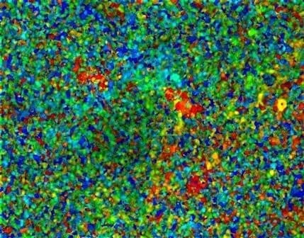
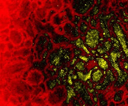
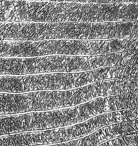
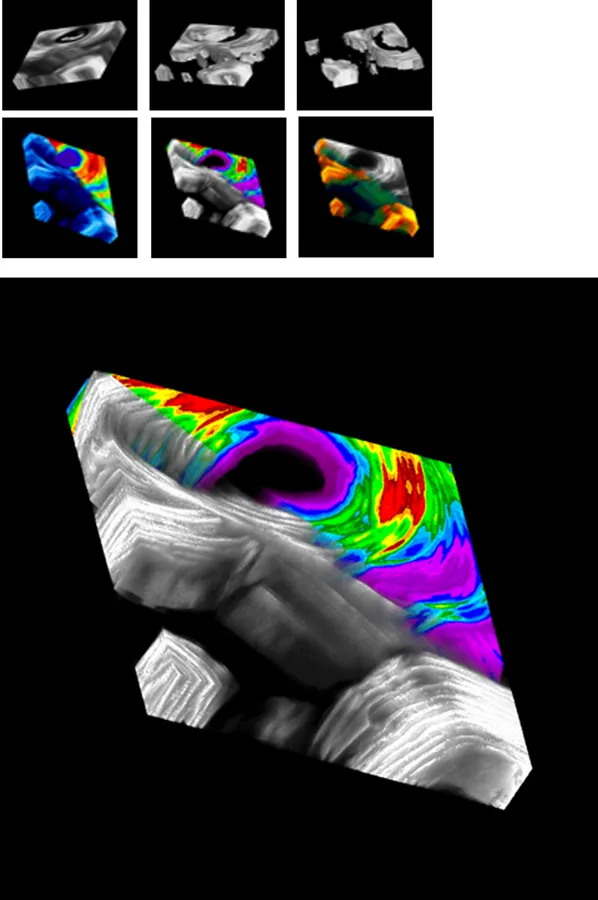
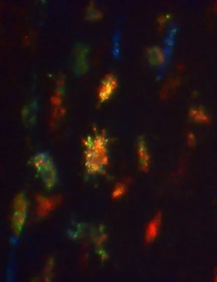
Image of bone microanatomy by portable confocal scanning optical microscopy. A novel portable Nipkow disk-based confocal microscope invented and patented by the artist was employed in the imaging of femoral bone from the famous “Lucy” discovered from fossil bearing deposits at Hadar, Ethiopia, approximately 3.0 m.y old. This image provides information about the degree of orientation of the cell spaces beneath the surface of the bone, which in turn can tell us about the way in which the bone was growing during childhood. Well oriented cells means that the surface was depositing bone during growth, while randomly oriented cells means that the surface was resorbing bone during growth; bone deposition, coupled with bone resorption, is how bones grow. Comparing the organization of bone cells between Lucy, belonging to the species Australopithecus afarensis, with other species of early human, can help us to understand changes in how bones grew over human evolutionary time. Original width 110 μm
You might also need